Tuesday, November 22, 2011

Breast Cancer Survival Rate - Stage 4 Breast Cancer


The breast cancer survival rate for Stage four breast cancer is significantly lower than for breast cancer detected at earlier stages.

Stage four breast cancer, or advanced breast cancer, has metastasized to other tissue such as bone tissue, lung tissue, or the liver. When breast cancer has overwhelmed the body's all-natural defenses and spread this far by the time the cancer is very first diagnosed, the 5-year survival rate drops to 16%-20% in the United States (American Cancer Society).

Up to 5% of white females in the U.S., and up to 9% of black ladies have advanced breast cancer spread to distant tissue at the time of very first diagnosis (SEER). This difference is often attributed to poverty and lack of health insurance.

In general, women who have advanced breast cancer at the time of diagnosis live roughly 18 months soon after diagnosis (median survival rate). Those who are nonetheless alive five years following their diagnosis of advanced breast cancer can live an extra three.five years (median survival rate) according to the American Cancer Society.

Given that this is the most deadly category of breast cancer, it is valuable to function closely with all the well being care providers. New remedies are getting developed all the time, and second, or even third opinions may perhaps give the patient much more information about newly discovered effective solutions.

Early detection is clearly the most significant element in breast cancer survival rates. Breast cancer detected at Stage 1 whilst it is still localized to the breast has a survival rate of 98%-100%, while metastasized breast cancer initial detected at Stage four drops down to 16%-20%.

Early detection procedures need to involve monthly self-examinations carried out at the exact same time each and every month. From age 20-40, wholesome women should certainly have clinical breast exams performed by their health care providers each and every 3 years. Immediately after age 40, the breast exams should be annually and ought to include things like a mammogram or similar process.

North American white girls have the highest rates of breast cancer in the globe, but the 5-year survival rate for all stages (Stage 1, Stage two, Stage 3, and Stage four) combined is 88% for the U.S. A current study identified European nations have lower 5-year breast cancer survival rates, with England at 77.8% and Ireland at 76.2% (Lancet Oncology).

The distinction in these survival rates is generally attributed to life-saving early detection.

What Stage 3 Breast Cancer Means


Stage three is classified into subcategories recognized as 3A, 3B, and 3C.

Stage three entails a mastectomy and lymph node removal as a standard of care. A joint approach of each chemotherapy and radiation is often recommended following surgery. Endocrine and targeted/biologic therapies might be options for therapy as nicely.

Stage 3A indicates
- Even although no tumour is observed in the breast, the lymph nodes beneath the arm include cancer cells and are stuck jointly, but there is no sign of cancer extend or
- The tumour is 5 cm or much less, the lymph nodes in the armpit contain cancer cells and are stuck to each and every other, but the cancer has not extend elsewhere or
- The tumour is more than five cm, the lymph nodes in the armpit consist of cancer cells and might be stuck jointly, but there is no further extend

Stage 3B indicates
- The tumour is fixed to the skin or chest wall, the lymph nodes may or may well not involve cancer cells, but there is no further extend

Stage 3C indicates
- The tumour could be any size and has extended to lymph nodes in the armpit and beneath the breast bone, or to nodes above or below the collarbone, but there is no further extend

Stage three breast cancer is typified by 1 of the following:
- A primary cancer that measures less than 5 cm (2 inches) in size and causes axillary (underarm) lymph nodes to be attached to each other or other structures
- A primary cancer that is greater than 5 cm (2 inches) in size and involves axillary lymph nodes
- A main cancer that is attached to the chest wall or skin

Breast cancer that has extended to the lymph nodes is frequently referred to as node-positive disease.

Effective therapy of stage 3 breast cancer needs each local and systemic therapy. Nearby therapy includes surgery and/or radiation and is directed at obliterating any cancer cells in or near the breast. Systemic therapy is aimed at at obliterating cancer cells all by way of the body, and may possibly consist of chemotherapy, hormonal therapy, or biological therapy. Systemic therapy may possibly be managed prior to surgery, which is named neoadjuvant therapy.

Monday, November 21, 2011

The Pathologist's Report of Breast Cancer


By this time you have received a diagnosis based about the biopsy findings. You know no matter if or not you've tumor. Your diagnosis was produced by the pathologist-the only member of your diagnostic team whom you most probably will not meet, consist of information on regardless of no matter whether or not cells are present, and the character from the cells which are observed under the microscope. A surgical pathology report may be within the form of the template or freely dictated, but either way it ought to include particular details. Don't just study the summary of the findings on the finish from the report.

Study and fully grasp the physique from the record. Look on the sample surgical pathology record at the end of this chapter. Note the separation of gross findings from microscopic findings. Though they are unique, each are very important towards the final diagnosis. The "gross pathology" might be dictated by a several pathologist from the 1 who signs the report, considering the gross pathology is done the day the specimen arrives in the pathology laboratory the tissue isn't study until it has been examined below the microscope by the pathologist who dictates the microscopic findings. The final surgical pathology report might not be typed and signed until days later.

The vital information you will need to glean from the gross pathology is the size, place, and character from the specimen muscle as a complete, and the size, place, and character from the tumor (if there is any) that may possibly be contained inside it. Do not confuse the two. The larger dimensions of the specimen as a entire are not the dimensions from the cancer. The size from the cancer has important implications for the "stage" from the tumor. If tumor is diagnosed within the specimen, the further description of the muscle, as set forth in the gross pathology, becomes essential, such as the location of the cancer within the specimen as aentire. For example, a pathologist frequently can make the diagnosis of inflammatory breast cancer when he or she sees, beneath the microscope, cancer cells in the lymphatic ducts of the skin-the "dermal lymphatics."

When the tumor is situated on the edge from the specimen and is cut through, a reexcision will be vital. The gross pathology has told the complete story. The microscopic pathology to come will simply confirm that the margin is very good. The color and also the consistency of the tumor inside the specimen are also relevant, in that they may well characterize the tumor. Right after dictating his or her findings, the pathologist who carries out the gross pathology will cut some of the tissue into modest pieces and put them into "cassettes," porous holders of the fragments. The cassettes are submerged in a fluid that preserves the tissue.

Please note that the pathologist cannot examine just about every cell in a core needle specimen, or inside the larger specimen of an open biopsy. He or she may well "bread loaf" the tissue by cutting it into slices like a loaf of bread and putting representative sections into cassettes. The remainder of the specimen is retained in jars containing preserving liquid, so that if there are any questions concerning the pathology, further muscle can be examined. Legally, the pathology department will need to maintain the preserved tissue for a specified period nothing will need to be thrown away at the time of the procedure. Suitable soon after an appropriate time, laboratory technicians prepare the preserved muscle further for the pathologist who will carry out the microscopic examination.

They location quite thin sections from the tissue on glass microscope slides, stain them appropriately, and cover them. The pathologist reads the slides below the microscope and dictates the "microscopic examination" portion from the record. The concluding summary provides the gist of the gross and microscopic findings. The most normal breast region cancer is called adenocarcinoma. The term is truly a composite: "adeno" describes the tissue of origin of the cancer "carcinoma" is honestly a fancy term for cancer. Thus, an adenocarcinoma is a tumor of glandular origin. And a breast area adenocarcinoma is really a breast area cancer of glandular origin.

The distinct muscle where the breast region cancer has originated is either inside the duct program from the breast (when it is recognized as ductal adenocarcinoma or, a lot way more typically, ductal carcinoma) or in the lobules (the part of the breast method precisely exactly where the milk is produced). Lobular carcinomas, when invasive, possess a life expectancy related to that of invasive ductal carcinomas. Despite the fact that they might have different characteristics, the two are subjected towards the very same therapy. (You should be conscious that invasive lobular carcinomas often are not visualized on screening mammograms basically because, it is believed, their outside edges have alot more tendrils and are not distinct.)

Each ductal and lobular carcinomas are treated differently when they are noninvasive than when they are invasive. A non invasive ductal carcinoma (otherwise known getting a DCIS, an acronym for ductal carcinoma in situ) consists of a distinctive treatment path from that of the lobular carcinoma in situ, LCIS. The record dictated from the pathologist is typed and submitted to him or her for approval and signature. Lastly, it is conveyed to you. You are in a position to see why it takes a few days to problem the pathology record, and a quantity of a lot even more days till you get the outcomes. Your physician might possibly wish to shorten the time involved by phoning the pathologist and acquiring an oral record.

But should you then get the report from your physician, who did not see the muscle beneath the microscope, it is undoubtedly feasible for error to creep into the transmission. Whenever you obtain the formal record, study all of it, not just the summary. If you do not understand the particulars, ask your physician to clarify them. Pathologists ought to know all concerning the tissue they are handling, such as the "natural history" (untreated history) from the tumor. The report will contain the answers to three big concerns: Do you have cancer? If so, what kind is it? And particularly, is it invasive or noninvasive? The pathologist's answers will have profound consequences for your remedy. If the margin is positive, the pathologist will need to be able to say how very good it's.

As you can see from Figure 6, the margin could be "grossly" positive (numerous, lots of cancer cells are there) or "diffusely" decent (only a relatively few cells can be observed). Obviously, when the yolk is off center, at the edge from the white of the egg, and is cut via, the margin will be referred to as grossly positive. Truly, if the tumor has been cut by way of, a major number of cancer cells will remain in the tumor bed (the remaining muscle in you). Even although it is essential for the team to know when the margin is grossly or diffusely excellent, the bottom line for you is that if the margin is recognized as positive, extra surgery-a reexcision of the margin of the tumor bed-have to be regarded as. If the pathologist's report proper right after your biopsy describes cells in the lymphatics of the skin, you possess a diagnosis of inflammatory breast cancer. If that specialized cancer is treated like plain old breast location cancer (POBC), the outcome could be disastrous.

Any suspicion of inflammatory breast location tumor need to be followed up, with a second opinion from an additional pathologist or oncologist if vital. As opposed to the remedy for POBC (surgical therapy, chemotherapy or hormonal therapy, and radiation), the sequence of remedy for inflammatory breast cancer is chemotherapy or hormonal therapy 1st, then surgery, and then radiation. If inflammatory breast tumor is treated getting a mastectomy on the outset, the cancer cells inside the dermal lymphatics are cut by way of on the time from the initial surgery and can spread all over the chest wall. Soon thereafter, tumor nodules can seem on the chest wall en curasse-covering the entire chest wall. This progression spells disaster for the patient.

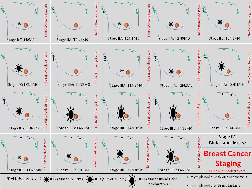
If chemotherapy or hormonal therapy can render the dermal lymphatics no cost of tumor, you will acquire two outcomes. Initial, the peau d'orange look from the breast region skin can disappear and, second, surgical remedy could be carried out safely. On the very same time, the systemic treatment affects the huge central mass of tumor and makes it much smaller, and subsequently surgically amenable to remedy. The pathologist has nonetheless one more role: to give the tumor a pathologic stage. This last and essential staging includes a substantial bearing on your future. The pathologist in no way creates formal remedy recommendations, because the pathologist is not a treating physician.

The remedy team makes therapy recommendations. Only the patient makes remedy decisions. The pathologist may possess a robust opinion about what the remedy ought to be, but it is not stated in the pathology report or in any formal setting in which the pathologist participates (for example, at a tumor board-about which you will hear a lot more in a moment). If the disease or tumor diagnosed is rare, the pathologist could comment appropriately inside the pathology record. Patients are frequently pleased to have their case presented to a tumor board. They picture that physicians with distinct specialties will especially meticulously evaluate their case. The operative word is quite cautiously. Optimally, the slides should really be presented by the pathologist assigned to the tumor board as well as by the presenting doctor. Presumably the pathologist has had time to evaluation the slides beforehand.

Similarly, the x-rays should really be evaluated prior towards the tumor board meeting and presented from the assigned radiologist. If the slides and x-rays are meticulously reviewed and presented, the role from the tumor board can be highly meaningful. Regularly, still, the films or slides are not present. Or the pathologist or radiologist is absent. Or the specialists have not had sufficient time to assessment the slides or films. Often the attending doctor is seeing the slides for the very first time. In such hit-or-miss circumstances, the board's recommendations may possibly not be definitely thoughtful or they could be biased in favor from the presenting physician.

Even if the tumor board is nicely organized and nicely prepared, the circumstance regularly is presented rather easily. The pathologist, the radiologist, and also the physicians on the board have small chance to think about the scenario, the patient is not seen, and the recommendations may be tainted from the presentation. There is no substitute for seeing and examining the patient and taking sufficient time to believe about the scenario appropriate after reviewing all the records, films, and slides. Tumor board recommendations are just that-recommendations. They need to never ever be accepted as definitive treatment choices. Your remedy team is responsible for explaining your treatment choices to you, and only you are in a position to make a decision what remedy you'll have.

Breast Cancer - Our Journey


I do a superb deal of speaking regarding breast well being and taking charge of one's life. As a motivational speaker you hope that your message reaches the spirit of your audience to take action with regards to their well being, particularly their breast well being. When it comes to our breast health, or any well being concern for that reality, as individuals we have to be willing to get to know our bodies and grow to be aware of any change from yesterday, last week, last month or last year.

Typically times our body will send us warning signs that we can heed or merely ignore. I believe in the adage "an ounce of prevention is worth a pound of cure." So when I meet ladies who tell me they have a household history of breast cancer and know they need to being performing factors to manage their breast well being but elect not to, due to the fact they prefer not to know if some thing is incorrect, I am left speechless. Breast cancer is the second leading trigger of cancer deaths, following lung cancer in ladies. If you had a blister on your foot would you not do something about it rather of letting it fester into a critical wound? Of course you would. That is why it is so valuable to follow the American Cancer Society recommendations to have annual mammograms right after the age of 40 (or younger for girls with a family history of breast cancer), have annual clinical exams and even do monthly breast self exams to know your body.

When my friend of thirty plus years told me she had been diagnosed with breast cancer last month my heart stopped. This was the 1st time somebody so close to me had heard those words "you have breast cancer." I asked if she had any indications of one thing wrong - an unfamiliar lump, skin discoloration, swelling or a strange secretion from the nipples. She told me this cancer was discovered through her mammogram and she was now scheduled for a lumpectomy the following week.

Following further discussion she shared that she had not had a mammogram in two years. At a concert on the National Mall grounds she and a stranger discussed a range of topics and 1 factor led to a different. She told the stranger she had not had a mammogram in two years. This stranger occurred to be a double mastectomy survivor and told my buddy to schedule her mammogram right away. I'm listening and thinking "have you not heard something I've been saying for the past 15 years?" Apparently not, but I'm glad God sent an angel to my buddy to get her moving.

She then disclosed to me that she had been diagnosed with DCIS (ductal carcinoma in situ) 3 years ago. DCIS is the most popular non-invasive breast cancer. It is non-invasive because it has not spread outside of the milk duct into the surrounding breast tissue. It is regarded as a Stage cancer, and remedy is removal of the cancer cells and surrounding margins.

I'm now floored as she continues to tell me she in no way stated anything since she did not want me to worry and make a huge deal about it. She is correct, in that I would make a massive deal about it. Getting been diagnosed with DCIS, her risk components had elevated for breast cancer to reoccur or generate a new breast cancer, which is exactly where she now finds herself. Delaying her mammograms was not a beneficial program of breast well being management.

The entire conversation made me realize that no matter how considerably we preach, teach or reach out to other people, the ultimate caretaker of one's well being is you. Persons will only tell you what they want you to know, and will only do what they are not afraid to do. If there is any drop of fear in their mind about a well being problem, that drop grows into a puddle, river, of ocean of fear that makes it harder each day to act on what 1 knows they must do.

Fear is a state of mind that creates a physical reaction of no action. The challenge we as a community face is to defuse the all-natural fear of hearing the word "cancer." More than 96% of women diagnosed early with no metastatic breast cancer (cancer that has not spread to other organs from the original web page) survive 5 years or way more. For the hundreds of thousands of ladies who proudly proclaim "I am a survivor" they are living testaments that there is life just after breast cancer. Do not let fear steal your life.

I explained to my dear friend that considering she has now been diagnosed with breast cancer, her daughter's threat variables have elevated. The girls in her household now have a greater danger element. This data desires to be shared, mainly because so a large number of girls believe there is no history of breast cancer in their family members. We have to be willing to speak openly about breast cancer in order to aid other people in our household manage their breast well being. The time for silence on this issue requires to finish.

My friend has just begun her journey with breast cancer. Her journey is now my journey, because she is my sister.

Saturday, November 19, 2011

Breast Cancer Survival Rate


When one talks of breast cancer and therapy, the secondary question is its survival rate. Survival rates give patients an thought of the extent of their cancer as well as the remedies that are out there to them. We typically hear of five-year survival rates for each stage of breast cancer, but what exactly is breast cancer survival rate?

Breast cancer survival can be described in the following approaches:

- Period of time : 5 or 10 years, that a woman lives right after diagnosis

- Risk of reoccurrence

- Risk of death when compared to others with the similar illness

The initial is the alot more widely used approach. Because the survival rate is frequently categorized according to stages, some points about the stages of breast cancer initially.

Stage cancer is the non-invasive sort. Cancer cells stay within the walls of the place where they are found. For Stage I, the tumor is invasive and is about two centimeters lengthy. Stage IIA cancer means that tumor is two-5 centimeters. With Stage IIB cancer, the tumor might be much less than two centimeters but a couple of axillary lymph nodes are affected. For Stage IIIA cancer, the tumor is extra than 5 centimeters or it has reached extra lymph nodes. Stage IIIB cancer is characterized by the tumor invading the breast skin, regardless of its size. Stage IV cancer is the most advanced form, exactly where the cancer cells have moved far from the breast and have infected other organs of the body as nicely.

Stages are also described as early (Stages -IIA), later (Stage IIB and III) and advanced (Stage IV).

When diagnosed with breast cancer it is the stages that will establish the therapy strategy.

Survival rates

In computing the survival rate, researchers take note of the percentage of women who survive for a precise period of time, say, five years, just after diagnosis of breast cancer. The present survival rates for all breast cancer stages are:

- 5-year survival rate - 86%

- Ten-year survival rate - 76%

Ladies with no metastatic breast cancer have a five-year survival rate of 96%, whilst those with metastatic breast cancer have a five-year survival rate of 21%

Here are the 5-year survival rates according to stage:

- Stage - 100%

- Stage I - 100%

- Stage IIA - 92%

- Stage IIB - 81%

- Stage IIIA - 67%

- Stage IIIB - 54%

- Stage IV - 20%

Keep in mind that these are estimates only. Some basically live longer than 7 years, depending on the medication and lifestyle alterations that they make. Immediately after 7 years survival rates reduce.

Other factors that have an effect on survival

Preliminary studies have been conducted regarding variables that can affect survival. There are promising results with respect to elements such as changing your diet program and lifestyle. Even though the results are not conclusive but, it still makes sense to preserve a healthy way of life. While there is no direct connection in between workout and elevated survival rate, studies showed that exercise improved the quality of life of survivors, such as greater self-esteem, improved mood and better sleep patterns. The same holds accurate for group psychological therapy. Getting able to express their feelings and support for other survivors had positive effects on their excellent of life. Smoking increases the risk of the spread of cancer, as there may well be metastasis of cancer from breast to lung.

The significance of early detection cannot be overemphasized. When detected early, the correct remedy can then be administered and once treated, there is much less danger for the cancer to spread or recur. That is why physicians and breast cancer advocates encourage standard testing and screening for all females. This is vital even right after remedy mainly because there is nonetheless a risk of recurrence.

Conduct a self breast exam monthly. If important, have clinical tests such as mammograms and MRI scans. Ask your doctor for a lot more information on breast cancer and search the web for answers from experts.

Breast cancer survival rates are mere estimates. Some patients basically live longer than 5 or 7 years. Modifications in diet regime and way of life can increase a patient's survival rate. Live a healthy way of life by consuming significantly more fruits, vegetables and fiber, and stay clear of alcohol intake.

Lung Cancer - Classification, Stages, Symptoms, Causes, Effects, Prevention, Detection and Treatment


Lung cancer is brought on by uncontrolled rapid growth of cells in tissues. This sort of cancer is most frequent and results in much more than a million deaths just about every year. This form of cancer is indicated by weight loss or coughing up blood or often going out of breath. It can be noticed on chest radio graph also referred to as CT Scan. The therapy that one gets depends on the stage that 1 is in. Treatment of cancer contain surgery, chemotherapy and radiotherapy.

CLASSIFICATION

Lung cancers are classified following studying under them microscope. Classification is required as diverse kind of cancer is treated differently. Massive portion of lung cancer are carcinomas - malignancies that grow from epithelial cells. Lung-carcinomas are categorized into two sorts: non -small and tiny-cell lung carcinoma. Non-smaller cell lung carcinoma and tiny cell lung carcinoma account for 80. 4% and 16. 8% frequency of lung cancer, respectively.

1. NON -Smaller CELL LUNG CARCINOMA

The non -small cell lung carcinomas are grouped together as their prognosis and management are exact same up to some extent. They are further classified into three kinds: squamous cell lung carcinoma, adenocarcinoma and substantial cell lung carcinoma. Squamous cell lung cancer originates near a central bronchus. They account for 25% of lung cancers. Adenocarcinoma begins in peripheral lung tissue. The situations of adenocarcinoma are a result of smoking. They accounts to 40% of non -modest cell lung cancers.

two. Smaller CELL LUNG CARCINOMA

This form of lung cancer is rare. It is at times referred to as "oat cell" carcinoma. Most of the occasions they originate from larger airways (primary and secondary bronchi ) and from there they grow at a rapid pace. This form of lung cancer if mostly associated with smoking.

SECONDARY CANCERS

These cancers are classified on the basis of webpage of origin like breast cancer but has spread to the lung. Majority of the lung cancers in kids are secondary.

STAGING OF LUNG CANCER

Lung cancer staging is made use of to asses the degree of spread of the cancer from its location of origin. It is an significant factor that determines the possible remedy of lung cancer. The degree starts from 1A to four, 1A being finest prognosis and 4 being worst.

SIGNS AND SYMPTOMS

Following are the symptoms of lung cancer: 1. Voice becoming hoarse. 2. Sudden loss of weight. 3. Feeling pain in chest region or abdomen. four. Difficulty in swallowing. 5. Loss of appetite. 6. Running out of breath. A number of of the symptoms mentioned above are non -precise. By the time they notice symptoms or signs, cancer has already spread from location of origin. Incredibly couple of people today with this cancer have signs at time of diagnosis, these cancers are noticed on routine chest radio graph.

CAUSES

The three key causes of cancer are: carcinogens (which is found in tobacco ), viral infection and ionizing radiation. If exposed, it causes adjustments to DNA in tissue lining the bronchi of the lungs. With much more and alot more tissues acquiring damaged, cancer develops.

1. SMOKING

Smoking is the most important cause of cancer. In one cigarette, there are 60 unique known kinds of carcinogens like radioisotopes and nitrosamine. Smoking is believed to trigger 80% of these type of situations. The danger is usually much less in non -smokers. The time that a person smokes proportionately increases the chances of this cancer. There has been situations that if a individual stops smoking, the damaged cells gradually gets repaired. In non-smokers, passive smoking is the main causes of lung-cancer. Passive smoking is 1 inhaled from one other individual smoking.

two. RADON GAS

The gas produced from breakdown of radium. This gas is colourless and odorless. Exposure to radiation ionize the genetic material, causing mutations that from time to time turn cancerous. Exposure to radon gas is the second big trigger of lung-cancer following smoking.

3. ASBESTOS

Asbestos is responsible for causing a number of cancer, one amongst them is lung cancer. In UK, asbestos accounts for two to three% of the total instances of this cancer.

4. VIRUS

Viruses are responsible for causing lung-cancer in animals. And research has shown of comparable prospective in humans.

five. PARTICULATE MATTER

Particulate matter has a direct link to lung cancer circumstances. The size and quantity of particles in air determines the risk of getting lung-cancer. If concentration of particles increases beyond 1%, then the probabilities of getting this increases by 14%.

PATHOGENESIS

Just like might possibly other cancer types, lung cancer is began by activation of ocnogenes or inactivation of tumor suppressing genes. Ocnogenes are those genes that make consumers far more vulnerable to cancer. Ocnogenes are produced from proto-ocnogenes, when the latter is exposed to certain carcinogens. In k-ras proto-oncogene, mutations takes place which are responsible for ten to 30% of lung adenocarcinomas. Tumor invasion, angiogenesis, apoptosis, cell profileration are regulated by the Epidermal growth factor receptor. Mutations and amplification of EGFR are normal in non -tiny cell lung cancer. The basis for remedy with EGFR-inhibitors are also supplied by Mutation and amplification of EGFR. Chromosomal harm can lead to loss of heterozygosity which can result in inactivation of tumor suppressor genes. Harm to four of these chromosomes:3p, 5q, 13 q and 17 p are prevalent in modest cell lung-carcinoma. The p53, which is a tumor suppressor gene, located on chromosome 17p is affected in most of the circumstances. c-MET, NKX2-1, LKB1, PIK3A and BRAF are also mutated or amplified. A variety of genetic polymorphisms are supplementary to this cancer. Some of them involve polymorphisms in genes coding for interleukin-1, cytochrome p450, apoptosis promoters such as caspase-8, and XRCC1, which is DNA repair molecule. Folks getting these polymorphisms are far more likely to create lung cancer on getting exposed to carcinogens. The study has revealed that MDM2 309G allele is a low-penetrant risk factor for creating this in Asians.

DIAGNOSIS

If a individual has reported symptoms that might recommend cancer related to lungs, then chest radio graph is performed in the very first step. The test reveals the widening of mediastinium, atelectasis and pleural effusion. Even if there are no radio graphic findings but the hint of this is high since of things like the individual getting heavy smoker with blood-stained sputum then CT-Scan could present the necessary information. If findings are unnatural in cells in sputum, then they multiplies the danger of this sort of cancer. Early detection can be completed by Sputum cytologic examination together with other screening examinations. The differential diagnosis for those patients who show irregularities on chest cardiograph look into cancer related to lungs along with non malignant illnesses. These consider infectious reasons like tuberculosis or pneumonia. The above mentioned illnesses can lead to lung nodules.

PREVENTION

Prevention, just like often, is superior than cure. Actions in this direction have been taken by might possibly countries by identifying carcinogens and banning them but tobacco, which is the major cause of lung cancer, is nonetheless frequent. Eliminating cigarette smoking is very first hand target in the prevention of lung cancer. Methods to lessen Passive smoking have also getting taken by banning smoking in public areas and workplaces. New Zealand has restricted smoking in open areas. A similar step is also taken by Chandigarh, India. Bhutan has criminalized smoking since 2005.

SCREENING

Screening is used to detect disease by carrying out medical tests when the patient is not showing any symptoms. Chest radio graph or computed tomography are the tests utilized for screening of lung cancer. But, results have shown, that screening tests for lung cancer seldom has shown any benefit.

Remedy

The therapy of lung cancer can be accomplished in following approaches, depending on the stage or degree of cancer:

1. SURGERY

If physicians have detected lung cancer, then CT scan and positron emission tomography are in most cases applied to check if the illness is placed and surgery can be done or it has moved to the point where performing surgery is not doable. Surgery can only be performed if spirometry reveals superior respiratory reserve, but if it is poor, then surgery is not attainable. Even surgery has a death operative rate of 4. 4% but that is because of patient's lung function and other elements.

two. CHEMOTHERAPY

Chemotherapy, along with radiation, is made use of to treat tiny cell lung carcinoma. Primary chemotherapy is also utilized in metastatic non -modest cell lung carcinoma.

three. RADIOTHERAPY

Radiotherapy, with chemotherapy, is given when patient is not fit to under go surgery. This type of high intensity radiotherapy is named radical radiotherapy. CHART (continuos hyperfractioned accelerated radiotherapy ) is refined version of this approach in which a high dose of radiotherapy is given for a brief period of time. When cancer affects a short section of bronchus, then brachytherapy is given.

EPIDEMOLOGY

Lung cancer is the most widely reported cancer. There are 1. 35 million instances each and every year and 1. 18 million deaths. Lung cancer develop amongst those who have a history of smoking over a long period of years i. e 50 years and above. In addition to smoking, passive smoking is also a factor that causes lung cancer. Even the emissions from factories, automobiles, power plants pose a threat to human health. Lung cancer is found to have a reciprocal impact with sunlight and UVB exposure. This is due to effect of Vitamin D, created in skin throughout exposure to sunlight.